Varicose veins are swollen, twisted and sometimes painful veins seen under the surface of the skin. Usually, they occur in legs; however, they may occur in other parts of the body.
How Varicose Veins Are Diagnosed
In most cases, doctors diagnose varicose veins by physical examination alone. And, in certain conditions, other tests such as Doppler Ultrasound and Angiogram may be done.
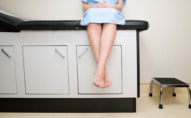
Physical Examination
Your doctor will look at your leg while you are standing or sitting with your legs dangling to check for swelling of the veins. Your doctor may also ask you about your symptoms, including any pain or itching in your legs.
Doppler Ultrasound
Doppler Ultrasound may be recommended to check the blood flow in your veins, if the valves of your veins are working properly or if there are any blood clots.
Doppler Ultrasound uses sound waves to make pictures of deep structures of your body. During this test, you lie on the examination table. Then, a small amount of warm gel is applied to the area of your leg being examined to prevent the formation of air pockets between your skin and the device. The technician then puts a hand-held device (transducer), about the size of soap bar, onto your skin and moves it over the whole area to be examined. The transducer transmits images of your veins to a monitor, so that your doctor can evaluate the state of your veins.
Angiogram
Angiogram is rarely recommended; however, your doctor may ask for it to get more detailed images of your blood vessels and the blood flow through them. Angiogram can help your doctor to confirm whether your problem is varicose veins or something else.
During this test, a dye is injected into your veins. Then, X-ray images of your leg, where the blood vessels are outlined by the dye, are done.